Mouse CSF1 Recombinant Protein (HEK293 Expression) — Tag Free
Price range: $198.00 through $3,600.00
 HEK293 × Chaperone-Assisted Technology
Produced using our proprietary Z-Active® platform — HEK293 mammalian expression with chaperone-assisted folding for proper disulfide bonding, N-linked glycosylation, and 5–10× higher bioactivity vs Leading Suppliers.
Overview
M-CSF, also known as CSF-1, is a four-alpha -helical-bundle cytokine that is the primary regulator of macrophage survival, proliferation and differentiation (1-3). M-CSF protein is also essential for the survival and proliferation of osteoclast progenitors (1, 4). M-CSF also primes and enhances macrophage killing of tumor cells and microorganisms, regulates the release of cytokines and other inflammatory modulators from macrophages, and stimulates pinocytosis (2, 3). M-CSF increases during pregnancy to support implantation and growth of the decidua and placenta (5). Sources of M-CSF include fibroblasts, activated macrophages, endometrial secretory epithelium, bone marrow stromal cells and activated endothelial cells (1-5). The M-CSF receptor (c-fms) transduces its pleotropic effects and mediates its endocytosis. M-CSF mRNAs of various sizes occur (3-9). Full length mouse M-CSF transcripts encode a 520 amino acid (aa) type I transmembrane (TM) protein with a 462 aa extracellular region, a 21 aa TM domain, and a 37 aa cytoplasmic tail that forms a 140 kDa covalent dimer. Differential processing produces two proteolytically cleaved, secreted dimers. One is an N- and O- glycosylated 86 kDa dimer, while the other is modified by both glycosylation and chondroitin-sulfate proteoglycan (PG) to generate a 200 kDa subunit. Although PG-modified M-CSF protein can circulate, it may be immobilized by attachment to type V collagen (8). Shorter transcripts encode M‑CSF that lacks cleavage and PG sites and produces an N-glycosylated 68 kDa TM dimer and a slowly produced 44 kDa secreted dimer (7). Although forms may vary in activity and half-life, all contain the N-terminal 150 aa portion that is necessary and sufficient for interaction with the M-CSF receptor (10, 11). The first 229 aa of mature mouse M-CSF shares 87%, 83%, 82% and 81% aa identity with corresponding regions of rat, dog, cow and human M-CSF, respectively (12, 13). Human M‑CSF is active in the mouse, but mouse M-CSF is reported to be species-specific.
Specifications
- Species: mouse
- Expression System: HEK293
- Sequence: Lys33-Glu262
- Tag: Tag Free
- Molecular Weight: 26.0 KDa
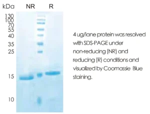
- Purity: > 95% by Tris-Bis PAGE;> 95% by SEC-HPLC
- Endotoxin: < 1 EU/μg of the protein by LAL method.
- UniProt/Swiss-Prot: P07141
- Available Sizes: 10 µg, 50 µg, 1 mg
Bioactivity
Measured in a cell proliferation assay using M‑NFS‑60 mouse myelogenous leukemia lymphoblast cells. The ED50 for this effect is 0.5‑3 ng/mL.
Formulation & Reconstitution
Formulation: 0.22 μm filtered solution of PBS, pH 7.4.
Reconstitution: 0.2 mg/mL in sterile PBS, pH7.4
Storage & Stability
Aliquot and store at -80°C. Avoid repeated freeze/thaw cycles.
Quality Assurance
Each lot is tested for purity (SDS-PAGE, SEC-HPLC), endotoxin levels (LAL method), and bioactivity. A Certificate of Analysis (COA) is provided with every order.
Safety Data Sheets & Documentation
Protocols & Support
Reconstitution Protocol
1. Briefly centrifuge the vial to collect lyophilized powder at the bottom.
2. Reconstitute in sterile deionized water or PBS to a concentration of 0.2–1.0 mg/mL.
3. Gently pipette up and down to dissolve. Do not vortex.
4. Aliquot and store at -20°C to -80°C. Avoid repeated freeze-thaw cycles.
Download Data Sheet
Complete product data sheet including batch-specific QC data, sequence information, and storage guidelines.
Download PDF Data Sheet